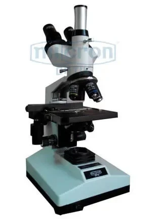
1. Viewing tube: As per Requirement
2. Head Rotatable at 360 degree.
3. Quadruple nose piece with positive click stops.
4. Seperate Coarse & Fine focusing Graduated to 1Div=.002mm.
5. With Mechanical stage for X&Y movement of slide.
6. Movable abbe Condensor N.A.1.25 with iris diaphragm with swingout filter holder.
7. Built in Illumination LED with intensity control. (Battery backup optional)
8. Steple Grey, chemical resistant, back-on finish.
9. Complete with Instruction Manual and dust cover.
10. Duly packed in styrofom packing.
EYE PIECES: WF 10X
OBJECTIVES:4X, 10X, 40X S/L, 100X S/L [Achromatic]
MAGNIFICATION: X40 – X1000
1. Wooden Carrying Case
2. Objectives: X20, X45 S/L, X60 S/L
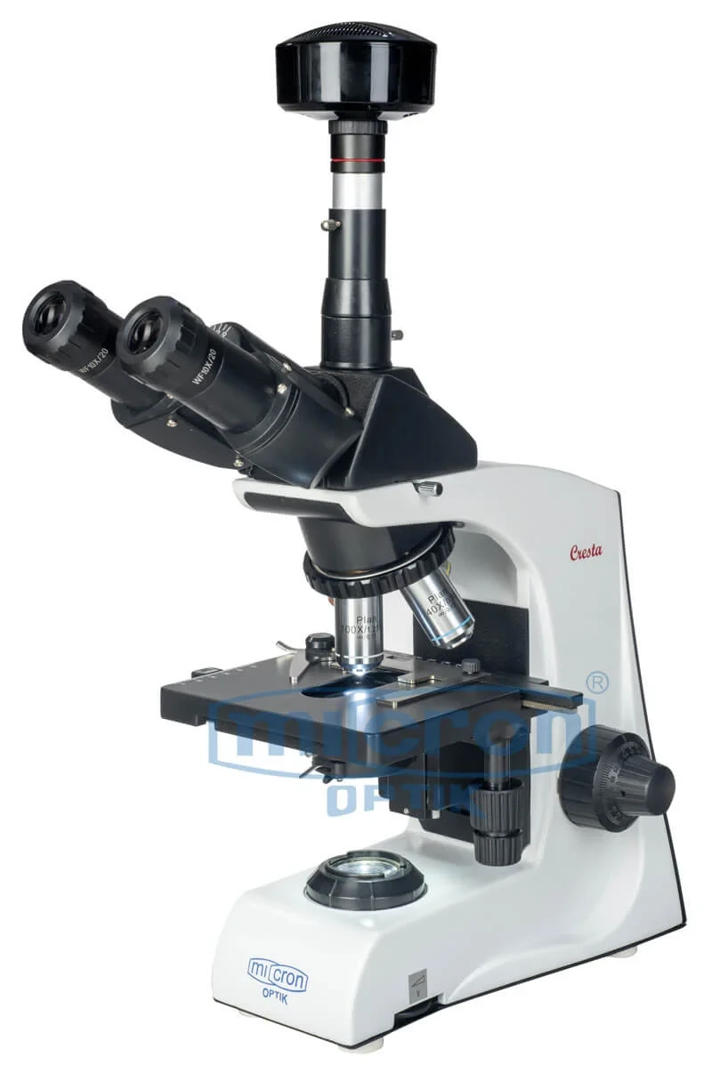
A trinocular microscope is similar to a binocular microscope consisting of two eyepieces and a third eyetube attached to its outer covering which acts as a microscopic camera.
The best trinocular microscope is designed with three different positions to ensure that the light is allowed at 360 degree angle for clear vision of the specimen. This helps in real-time viewing of the specimen. Full size trinocular microscopes are generally set at 100% to make fluorescence photography an easier task.
Micron's TRINO MINI brings the power of trinocular documentation to a compact and affordable platform, ideal for pathology labs and educational institutions that need to capture images and videos. The viewing head is rotatable 360 degrees and is available in monocular, binocular, and trinocular configurations. This model is widely recognized as one of the best entry-level trinocular microscopes for professionals who need to integrate digital imaging into their workflow. The quadruple nosepiece with positive click stops ensures reliable objective changes. Separate coarse and fine focusing knobs, graduated to 1 division = 0.002mm, provide precise control for critical focusing. Micron has equipped this model with a mechanical stage for smooth X and Y slide movement, which is essential for systematic specimen examination. For small pathology labs seeking a top rated trinocular microscope for documentation and consultation, the TRINO MINI is an excellent choice. The movable Abbe condenser N.A. 1.25 with iris diaphragm and swing-out filter holder gives users the ability to optimize contrast for different specimen types and for photography.
Trinocular Viewing Head: The third eyetube allows for the permanent attachment of a digital camera, enabling image and video capture for documentation, consultation, and teaching.
360-Degree Rotatable Head: The entire viewing head can be rotated 360 degrees, allowing for collaborative viewing and flexible positioning for photography or teaching setups.
Separate Coarse and Fine Focus: Independent coarse and fine focusing knobs provide precise, graduated control (0.002mm divisions), essential for achieving sharp focus for digital image capture.
Mechanical Stage with X-Y Control: The built-in mechanical stage allows for smooth, precise slide movement, enabling systematic specimen examination and easy tracking of specific fields for photography.
Oil Immersion Capability (100X): Comes complete with a 100X S/L oil immersion objective, providing 1000X magnification necessary for capturing high-resolution images of bacteria and cellular details.
Variable Intensity LED Illumination: The built-in LED light with intensity control provides a bright, cool, and consistent light source, ideal for both visual observation and digital photography.
Movable Abbe Condenser: The N.A. 1.25 Abbe condenser is focusable and includes an iris diaphragm, allowing the user to optimize contrast and resolution for the camera sensor.
Light Path Selector: The trinocular head features a light path selector, allowing the user to direct 100% of the light to the eyepieces, 100% to the camera, or split it 50/50.
Digital Pathology and Telepathology: Ideal for capturing and sharing digital images of slides for remote diagnosis, second opinions, and archiving in a digital pathology workflow.
Medical and Graduate Education: Perfect for connecting to a monitor or projector in a lecture hall, allowing an entire class to view a specimen simultaneously through the camera.
Clinical Case Documentation: Excellent for documenting interesting or rare cases by capturing high-quality images to include in patient records, case reports, or publications.
Small Clinic Pathology Labs: A compact and affordable solution for small labs that need to occasionally capture images for consultation or patient education without a large capital investment.
Quality Assurance in Industry: Can be used in manufacturing QA/QC departments to capture images of defects or features for reports, analysis, and process control documentation.
Research Data Collection: Suitable for researchers who need to capture high-quality micrographs of their specimens for use in presentations, papers, and grant proposals.
Veterinary Diagnostic Imaging: Allows veterinary pathologists to capture images of animal tissue samples for consultation with specialists or for educating pet owners.