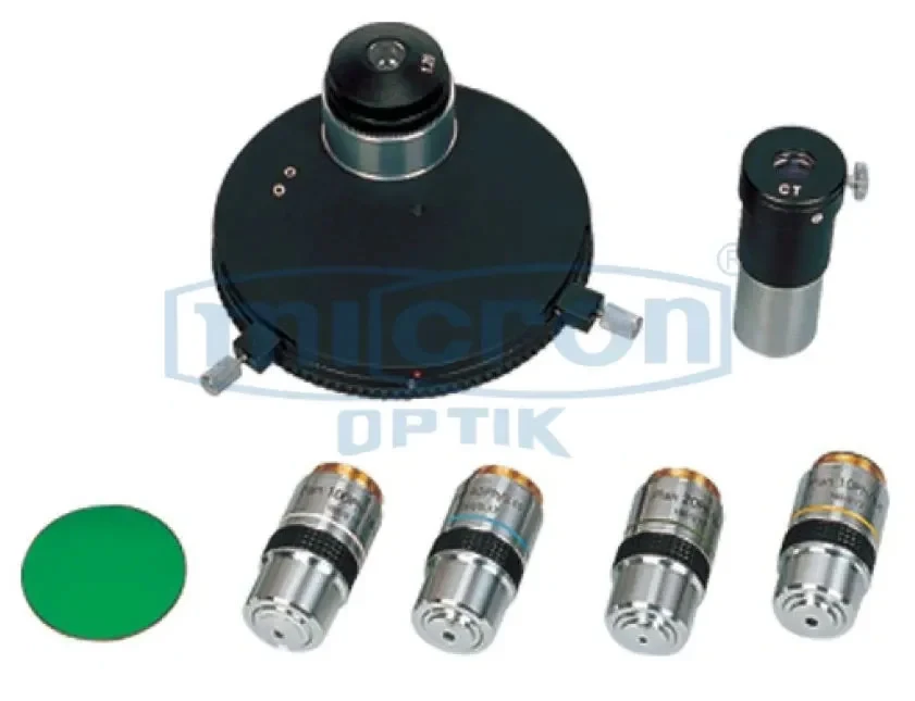
DARK FIELD ATTACHMENT
Detailed Dark Field Attachment Specifications: Type: Dry dark field condenser or oil dark field condenser (specify). Design: Paraboloid or cardioid condenser type, designed to produce a hollow cone of light. Compatibility: Fits into the standard substage condenser mount of most compound microscopes (e.g., 37mm dovetail). Numerical Aperture: Typically N.A. 0.80-0.95 for dry, up to 1.20-1.40 for oil immersion. Central Stop: Contains a central light stop to block direct light. Material: Constructed from black anodized aluminum or brass with high-quality optical glass. Included: Condenser unit, central stop, and sometimes immersion oil. Usage: Replaces the standard Abbe condenser. Requires a bright light source (e.g., 30W halogen or higher). Not suitable for low magnifications (below 10X objective) or for specimens that are too thick.
Micron's Dark Field Attachment is a specialized optical accessory designed to enhance the contrast of unstained, transparent specimens. Dark field microscopy works by illuminating the specimen with a hollow cone of light, causing only the light scattered by the specimen to enter the objective. This results in a bright specimen appearing against a dark, almost black background. This technique is widely considered one of the best methods for observing live, unstained microorganisms and fine details in transparent materials.
This attachment is designed to be easily fitted to a standard brightfield microscope, typically by replacing the standard Abbe condenser. The dark field attachment creates the hollow cone of light necessary for the technique. This is one of the best and most cost-effective ways to add dark field capability to an existing laboratory microscope. It is ideal for observing living organisms, such as bacteria, protozoa, and algae, without the need for staining, which can kill or alter the specimen. It is also excellent for examining fine structural details in diatoms, fibers, and other transparent materials. Micron's dark field attachment is a simple yet powerful tool that dramatically expands the capabilities of a standard brightfield microscope. For educational labs and clinical settings, this is a top rated accessory for observing live, unstained specimens.
Micron's Dark Field Attachment is especially useful for viewing motile microorganisms in their natural state, enabling researchers, students, and clinicians to study behavior, morphology, and movement patterns with improved visibility. The enhanced illumination method also supports examination of thin fibers, crystal fragments, insect parts, and other transparent or semi-transparent materials where edge contrast is essential. In educational settings, it provides an engaging and high-contrast visual experience that helps learners better understand specimen structure and optical principles.
The attachment is manufactured using durable optical-grade components to ensure consistent performance and long-term reliability in laboratory and teaching environments. Its design supports smooth installation and compatibility with a wide range of microscopes, allowing institutions and professionals to upgrade their existing equipment without investing in a completely separate dark field microscope system.
High-Contrast Imaging of Live Specimens: The dark field attachment creates a bright specimen image against a dark background, ideal for observing live, unstained microorganisms without killing them.
Simple Retrofit for Brightfield Microscopes: Designed to easily replace the standard Abbe condenser on most compound microscopes, providing an instant dark field capability upgrade.
Reveals Fine Structural Details: Excellent for visualizing fine details in transparent specimens such as diatoms, flagella, spirochetes, and other structures invisible in brightfield.
Ideal for Clinical and Educational Use: A perfect tool for clinical labs to quickly screen for spirochetes or for educational labs to observe live pond water organisms.
No Specimen Staining Required: Allows for the observation of living specimens in their natural state, avoiding the artifacts and killing effects of chemical stains.
High-Quality Optical Construction: Made from black anodized aluminum and high-quality optical glass to minimize glare and maximize light efficiency.
Compatible with Various Objectives: Works effectively with 10X, 20X, 40X, and 100X objectives (oil immersion for 100X) for a wide range of magnifications.
Durable and Easy to Use: A robust, low-maintenance accessory that is simple to install and focus, providing immediate benefits with minimal setup.
Microbiology and Bacteriology: Essential for observing live, unstained bacteria, particularly for detecting the presence of thin, hard-to-stain organisms like Treponema pallidum (syphilis).
Live Specimen Observation in Education: Perfect for high school and university biology labs to observe live protozoa, algae, and other microorganisms in pond water without staining.
Clinical Diagnostics: Used in clinical labs for rapid screening of wet mounts for certain pathogens, such as identifying motile spirochetes or for examining urine sediment.
Marine and Freshwater Biology: Ideal for examining live plankton, diatoms, and other small aquatic organisms in their natural, unstained state for research and monitoring.
Materials Science (specific applications): Can be used to examine surface defects or internal structures of transparent materials like glass, polymers, and crystals.
Forensic Science: Useful for detecting and examining trace evidence such as small fibers, hair fragments, and glass shards that are transparent or difficult to see in brightfield.
Quality Control in Industry: Applied in some QC settings to inspect transparent films, coatings, and liquids for the presence of small particles or defects.